


Papilloma of the neck is one of the manifestations of an infectious disease caused by human papillomavirus. Refers to benign skin formations.
Causes of Papillomas in the Neck
There is one etiological reason why papillomas start on the neck or other areas of the human body - infection with the human papillomavirus (HPV), which is a member of the Papovaviridae family. There are more than 100 serotypes of this pathogenic agent, each of which is responsible for the formation of a different clinical picture of the disease (papilloma, condyloma, warts - these concepts are synonymous, different names are associated with local characteristics).
The main ways of transmission are contact-household and genital organs (condyloma of the perianal region). The virus can penetrate the skin only in the presence of micro-lesions or open wounds, in other cases it can not cross the protective barrier of the skin.
Information about the pathogen
- It has a high prevalence, regardless of gender (however, it is more common in women than men), age or region (according to some sources, 2/3 of the planet is infected with this virus).
- contains double-stranded, twisted-strand DNA that can integrate into the human genome.
- Some strains of infection are associated with a high carcinogenic risk, especially in the case of permanent damage. Neck papillomas are caused by non-oncogenic strains of the virus. The virus goes through two main stages in the
- division process. In the first stage it is in episomal (free) form and in the same period the main division of the viral particle occurs. This stage is reversible (long-term remission occurs after treatment). In the second - integrative - stage, the virus is implanted in the cell genome (the first step towards cell degeneration and malignancy). The first stage is transitional and passes relatively quickly, while the second is hidden and explains the existence of trains.
- acts on the base layer of the epidermis where the virus multiplies. In the rest of the layers the pathogen can be retained but not divided. Provided the virus is in the embryonic layer as it grows, the normal differentiation of cells in all layers of this area is violated, especially at the level of the adhesive layer.
- has a tendency to move the body asymptomatically (from a few months to a year). It is rarely possible to identify a specific moment of infection - this is why treatment is started during the period of intense clinical manifestations and not at the first vague signs.
Predisposing factors
- Lack of hygiene. Since the virus can maintain vital activity in the outdoor environment for a long time, it is necessary to follow the rules of personal hygiene in public places (swimming pool, bath, gym).
- Traumatic skin lesions. Microcracks or scratches on the skin (for example, caused by scratching the neck with a shirt collar) are sufficient for the virus to penetrate.
- Immune system dysfunction. With immunodeficiency of any genesis, favorable conditions arise for the development of any infection. For example, frequent colds and infectious diseases cause a weakening of the immune system and the appearance of papillomas on the skin.
- Self-harm during skin tightening.
- Systemic lifestyle disorders (stress, physical activity, poor diet). These factors affect the work of all metabolic processes in the body and cause a reduction in the barrier function of the skin.
- Environmental factors that affect the body's reduced defenses (hypothermia, excessive exposure to ultraviolet light).
External manifestations of the disease
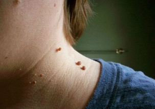
Cervical papillomas look like this in the photo:
- The growth is most often located at the broad base and protrudes significantly above the skin surface. Less often, the base of the papilloma is represented by a thin foot (in this case, the formation takes place in a hanging position). In the second variant, the risk of injury is much higher.
- The boundaries of education are equal and clear.
- The color is no different from the surrounding skin. In rare cases, it may be somewhat paler or darker than the surrounding tissues.
- The surface is often smooth, smooth. Sometimes it is possible for growths to form on the upper part of the papilloma, making its surface ribbed.
- diameter varies widely - from 1-3 mm to several centimeters (small diameter papillomas are more common).
- Location around the neck (back, side). Sometimes the face interferes.
Typically, there are multiple lesions located along the folds of skin.
In very rare cases, papillomas on the neck can become malignant, or turn into a skin tumor. This can occur as a result of infection with an oncogenic HPV strain.
Signs that may indicate a malignant transformation include:
- color change and heterogeneity (polymorphism);
- border change (blurring, loss of definition);
- appearance of asymmetry (it is impossible to get two equal parts when passing through the conditional middle line of formation);
- Intense growth;
- bleeding or ulceration (nonspecific symptom, as it is characteristic of simple lesions of the neoplasm);
- itching, burning, peeling;
- casting is formed (small daughter formations around the center).
Having such signs does not necessarily mean degeneration of the papilloma, but it does mean that you need to see a doctor and make a differential diagnosis to find out if we are talking about a common inflamed mole or skin cancer.
How to get rid of papilloma on the neck
Treatment of papillomas on the neck is carried out only in a complex way, simultaneously acting on the pathological focus of the skin and the pathogen itself in the blood.
There are several ways to fight:
method |
Description |
Medications |
The use of cytostatics, immunomodulators, is intended to prevent replication of a viral agent in the affected area and to reduce its concentration in the blood. Some drugs (keratolytics) are used topically to directly destroy skin growth (cauterize and cause tissue necrosis). |
Physical methods |
Cryodestruction, laser therapy, electrocoagulation. Their purpose is to get rid of papillomas on the neck as well as on other parts of the body. These methods allow you to restore the aesthetic appearance in open areas and remove the viral reservoir - skin neoplasms themselves, but they do not completely remove the virus from the body. |
Combination Therapy |
combines the two previous options and is therefore the most effective. |
Treatment of papillomas with folk remedies (for example, celandine juice) is ineffective and often dangerous, in any case, it is a prerequisite to consult a doctor.
Physical methods of destruction
It is possible to effectively reduce formations by using the following physical methods:
method |
Description |
Local action with concentrated acid solution |
Zinc chloropropionate 1. 5% solution is used in 50% 2-chloropropionic acid, a combination of nitric, acetic acid, oxalic, lactic acid and copper nitrate trihydrate, etc. . . . The agent is applied pointwise with a spatula until the color of the formation changes slightly (as soon as this has happened, further use should be stopped immediately). For a complete cure of papilloma, on average, 1-2 treatments are needed. |
electrocoagulation |
With the help of a special electric knife the point excision of the formations is done without affecting the underlying tissues (there is minimal impact on healthy skin cells). The method is most convenient when the formation has a long stem and a small size. |
Cryodestruction |
The focus is on liquid nitrogen, ultra-low temperatures leading to tissue necrosis. It is good that this form of cleaning is done on a wide base. The nitrogen action time is chosen by a specialist (1-5 minutes). After moxibustion, a burn is formed that heals in an average of 10 days. |
Laser removal |
The most modern and delicate approach to removing growths in prominent areas such as the neck. Has the most positive reviews. In continuous mode for 5 seconds to 3 minutes with the help of a light guide they act on the focus. The treatment period is much shorter than with other methods (5-7 days). The technique is associated with minimal trauma to the surrounding tissues, due to the high accuracy of the action. |
Classical surgical removal (excision with a scalpel) |
Used extremely rarely, only with large lesions or suspected malignant tumors. The reason is that the lesion is often multiple, scattered around the neck, and too small for excision, in addition, after surgical excision, scars may remain, which in itself creates a cosmetic defect. |